
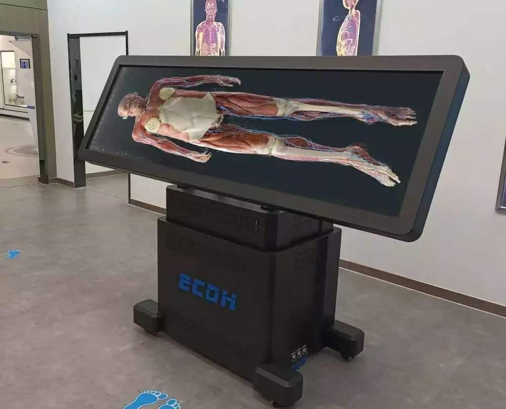
Medical education relies heavily on direct spatial understanding of human anatomy. For centuries, physical cadaver dissection has been one of the primary methods for teaching human anatomy. However, modern medical schools face persistent administrative and environmental challenges. Procurement costs remain consistently high. Specialized laboratory ventilation systems are mandatory to control hazardous preservative vapors.
Furthermore, a growing global shortage of physical specimens severely limits student access to hands-on dissection opportunities. This approach also addresses ethical concerns surrounding body donation and hazardous chemical storage. To address these urgent limitations, academic institutions are actively turning toward high-definition medical visualization. Digital technology now bridges the gap between traditional dissection and modern classroom needs.
This transition provides scalable, repeatable, and non-destructive learning opportunities. It empowers universities to train future healthcare professionals with advanced interactive tools. This helps institutions provide more consistent access to anatomical learning resources.
The Role of a Virtual Dissection Table in Modern Curriculums
Traditional physical dissection is a single-use process. Once an incision is made, the specimen cannot be restored to its original state. A virtual dissection table reduces this limitation by allowing students to repeatedly explore digital anatomical models. Students can manipulate high-resolution 3D anatomical models repeatedly without consuming physical specimens. They can zoom, rotate, and dissect layers of tissue without consuming valuable physical resources. This repetitive virtual practice builds tactile confidence before students ever enter a clinical setting. This active exploration fosters a much deeper understanding of complex spatial relationships inside the human body. For instance, tracing deep cranial nerves or isolating tiny vascular pathways becomes an intuitive process.
In addition, these digital platforms substantially lower long-term institutional expenses. They can reduce the constant, recurring expenses associated with importing, preparing, storing, and disposing of physical cadavers. A virtual dissection table also offers a highly safe, toxin-free learning environment for students. It reduces exposure to chemicals commonly associated with traditional cadaver preservation, such as formalin. Educators can preset specific teaching paths, custom-annotated structural pins, and interactive test questions. This keeps students highly engaged during self-guided study sessions. The technology acts as an essential, scalable bridge, allowing modern schools to significantly expand their medical training capacity.
High-Precision Dataset Engineering and Tomographic Images
The clinical and educational value of digital anatomy depends entirely on spatial accuracy. Leading educational solutions utilize ultra-high-definition human tomographic image datasets. These datasets provide detailed anatomical structures with high-resolution imaging capabilities. For example, some models utilize a male dataset containing 17,000+ continuous cross-sections. They also incorporate a female dataset with 16,000+ continuous cross-sections. Every single slice maintains a precise 0.1 mm layer thickness. The individual voxel size within these digitized datasets measures exactly 0.0384 mm × 0.0384 mm × 0.1 mm. This level of resolution helps capture detailed anatomical structures for educational visualization.
These high-precision datasets reconstruct nine major human systems. Students can observe and manipulate more than 6,000 distinct, accurately labeled anatomical structures on screen. To connect anatomical theory with clinical practice, the platforms integrate extensive real-world medical imaging. This includes over 1,700 real CT and MRI scans mapped directly to the specimens. Learners can match sectional tomographic specimen views directly with clinical imaging. This feature helps students better understand the relationship between anatomical structures and medical imaging. The integration of high-precision datasets ensures that students build correct, three-dimensional mental maps of human organs.
Expanding Beyond the Screen with 3D Printing Technology
Modern medical visualization does not end with interactive flat screens. Advanced medical programs combine digital screens with physical replicas. High-precision digital human data serves as the blueprint for full-color, multi-material 3D printing. This hybrid approach offers a tactile element that complements screen-based learning. It gives students a tangible feel for organ weight, spatial depth, and tissue flexibility. This hybrid training model bridges the gap between digital models and real physical specimen handling.
The manufacturing process uses specialized 3D printing equipment to replicate human specimens. These printers utilize 3D inkjet printing and light curing technology. They operate with 12 distinct material channels. This allows for the simultaneous printing of hard and soft components in a single build. The printers feature up to 3840 piezoelectric spray holes. They maintain a highly efficient output, spraying up to 4 liters of photopolymer material per hour.
The resulting physical specimens closely resemble real biological tissues. For example, a printed model can accurately show both rigid bone structures and soft, flexible blood vessels. This combination provides hand surgeons, neurosurgeons, and orthopedic specialists with highly realistic physical models. These physical models are ideal for surgical planning, professional training, and doctor-patient communication.
Choosing the Right Interactive Hardware for Medical Labs
When selecting digital anatomy systems, institutions evaluate hardware configurations carefully. Educational spaces vary significantly, demanding flexible equipment setups to fit diverse lecture halls. Standard hardware sizes for a virtual dissection table include 55-inch, 86-inch, and 98-inch all-in-one machines. These displays feature a native resolution of 3840×2160 pixels, supporting full 4K output. They also offer a static contrast ratio of 5000:1 and up to 500 cd/m² brightness. This ensures crisp, clear visualization even under bright laboratory lighting.
For large group laboratories, specialized tables feature a broad 88-inch screen. This configuration offers a 3840×1080 resolution. It includes high-performance computer processors, dedicated graphics cards, and fast solid-state drives. These high-spec components render complex 3D structures in real time.
The hardware supports multi-touch inputs, allowing multiple students to collaborate simultaneously on a single specimen. The system is also compatible with active stereoscopic projection, enabling a highly immersive 3D viewing experience for large classrooms. Furthermore, dual-language capabilities (English and Chinese) ensure international usability across global medical departments. Institutions evaluating digital anatomy solutions can review DIGIHUMAN’s virtual anatomy systems for available configurations and applications.
